Knee Anatomy
An Overview on Knee Anatomy
The human knee is a complex joint with many components making it vulnerable to injury. The anatomy of the knee allows the joint to flex, extend, twist side to side and handle a significant load of stress daily. Dr. Nikhil Verma, orthopedic knee specialist treating patients in the Chicago, Westchester, Oak Brook and Hinsdale, Illinois area, is highly trained and skilled on knee anatomy and the common knee injuries experienced by the general population, weekend warriors and professional athletes.
Anatomy of the Knee
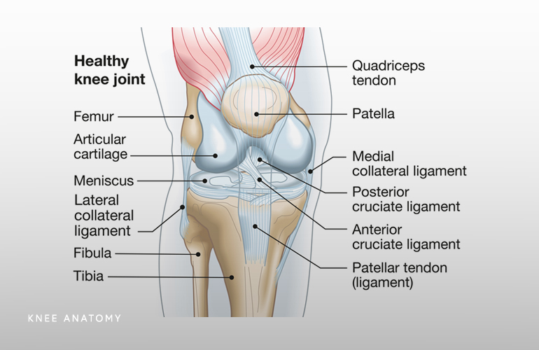
The knee joint is the largest joint in the body. It is made up of four main structures- bones, cartilage, ligaments and tendons. Three bones connect to form the knee joint. These bones include:
- Thighbone (femur)
- Shinbone (tibia)
- Kneecap (patella)
The anatomy of the knee also includes the articular cartilage, an extremely hard and smooth substance that decreases knee friction and helps the knee bones glide smoothly as the knee bends and straightens. Two wedge-shaped pieces of meniscal cartilage serve as “shock absorbers” between the femur and tibia, which help to decrease load on the articular cartilage and prevent development of arthritis.
The three bones are connected to other bones by ligaments. These ligaments act like strong ropes to hold the bones together and provide knee stability. These ligaments include:
- Anterior cruciate ligament (ACL): This ligament travels from the anterior of the tibia to the posterior of the femur and prevents the tibia from moving forward. This is one of the most important structures in the knee and is the most commonly injured in twisting movements.
- Medial collateral ligament (MCL): This ligament is a band that runs between the inner surfaces of the tibia and femur and prevents the knee from collapsing inwards.
- Posterior cruciate ligament (PCL): This ligament travels from the posterior of the tibia to the anterior of the femur and wraps around the ACL. It is the largest and strongest ligament in the knee and is infrequently injured. It prevents the tibia from moving backwards on the knee.
- Lateral collateral ligament (LCL): This ligament is located on the outside of the knee on the outer surface of the femur and fibula. It resists impact from the knee’s inner surface and prevents the knee from collapsing outwards.
The collateral ligaments control the sideways motion of the knee and brace it against unusual movement. The cruciate ligaments control the back and forth motion of the knee.
The remaining structures of knee anatomy are the tendons and muscles. The patellar tendon stretches from the patella to the shinbone. The quadriceps tendon connects the front thigh muscles to the patella. These muscles allow you to extend your knee and make it straight.
The anatomy of the knee is complex and involves numerous moving structures. Any of these structures can become injured from a fall, sports collision, overuse, a degenerative condition or accident.
Common knee injuries include:
- Knee fractures
- Knee dislocation
- Patella dislocation
- Knee tendon ruptures
- Ligament sprains and tears
Many of the common knee injuries can be treated by a non-surgical approach. If surgery is needed, Dr. Verma and his sports medicine team will determine the proper surgical technique to alleviate pain, improve knee stability and return the anatomy of the knee to its proper condition.
If you would like more information on knee anatomy and common knee injuries, contact Dr. Nikhil Verma, orthopedic knee specialist serving the Chicago, Westchester, Oak Brook and Hinsdale, Illinois communities.

